| 17 مايو 2026م
عن الجامعة
الشؤون الأكاديمية
البحث العلمي
الموارد والخدمات
English
مركز دارس
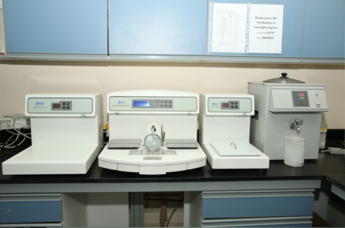
وحدة المناعية و الأنسجة
عن الجامعة
الشؤون الأكاديمية
البحث العلمي
الموارد والخدمات
English
مركز دارس
وحدة المناعية و الأنسجة
عن الجامعة
الكليَّات والأقسام
البحث العلمي
الموارد والخدمات
English
إغلاق
عن الجامعة
من أقوال جلالة السُّلطان المعظّم
مجلس أمناء الجامعة
كلمة رئيس الجامعة
الإدارة العُليــــا
مجالس الجامعة
الدَّوائر والوحدات الإداريَّة
مجتمعُـــنا
إشاداتٌ وإنجازات
وظائف شاغرة
الاتِّصــال بنـا
إغلاق
الكليات والمعاهد
مكتـب نائـب الرَّئيـس للشُّـؤون الأكاديميَّـة
الدِّراسـات العُليــا
مكتب مساعد نائب الرئيس للخدمات الأكاديمية
عمادة التعلم الإلكتروني
عمادة القبول والتسجيل
نظام إدارة جودة التدريس والتعليم
الصفات المُميِّزة لخرِّيجِ جامعةِ نزوى
إغلاق
البحث العلمي
مركز دارس
الخدمات الاستشارية وتوطين الابتكار
مركز أبحاث العلوم الطبيعية والطبية
مكتب نائب الرئيس للبحث العلمي
الكرسي الوطني لعلوم المواد والمعادن
مركز الخليل بن أحمد الفراهيدي للدِّراسات العربيَّة والإنسانية
كرسي اليونسكو لدراسات الأفلاج وعلم المياه الاجتماعي
إغلاق
الموارد والخدمات
معهد الضاد لتدريس العربية للناطقين بغيرها
معهد التَّعلُّم مدى الحياة
مركز نظم المعلومات
خريطة الجامعة
عمادة التعلم الإلكتروني
دليل الموظفين
خريطة الموقع
البريد الإلكتروني للموظفين
البريد الإلكتروني للطـُّـلاب
مكتبة الجامعة
مركز مهارات الكتابة
مركز تعزيز مسالك التعلم
مركز ريادة الأعمال
المنظومة التعليمية
عمادة القبول والتسجيل
منظومة إدارة المساقات (موودل)
عمادة شؤون الطُّلاب وخدمة المجتمع
مركز الإرشاد والمتابعة الأكاديمية
الصحة و السلامة
اِستمع